
Operative Spinal Cord Anatomy The Neurosurgical Atlas
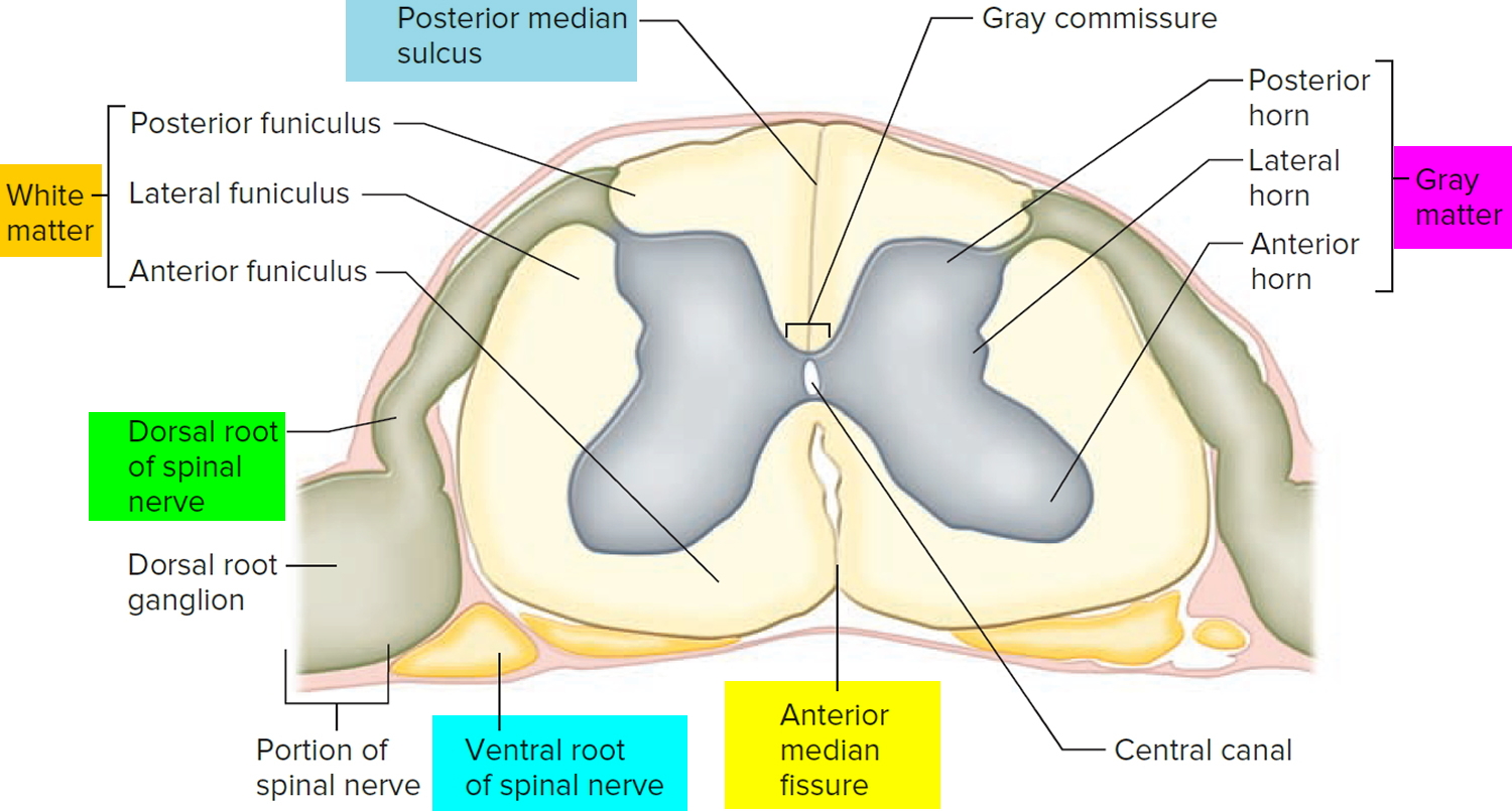
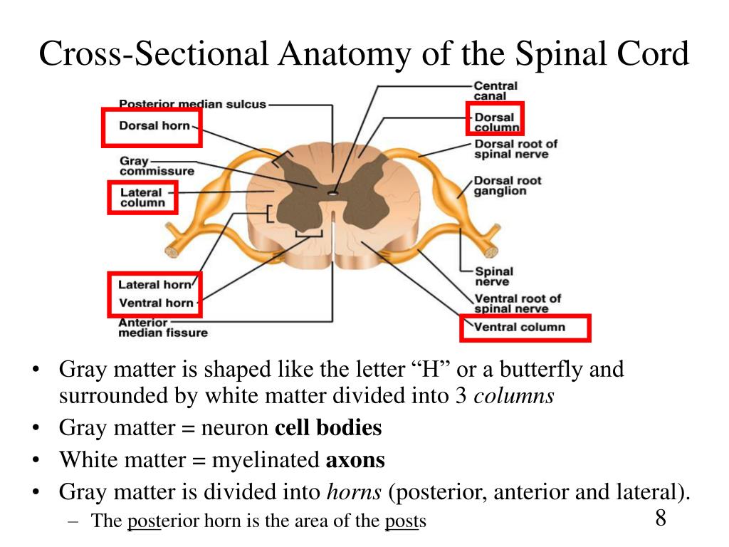
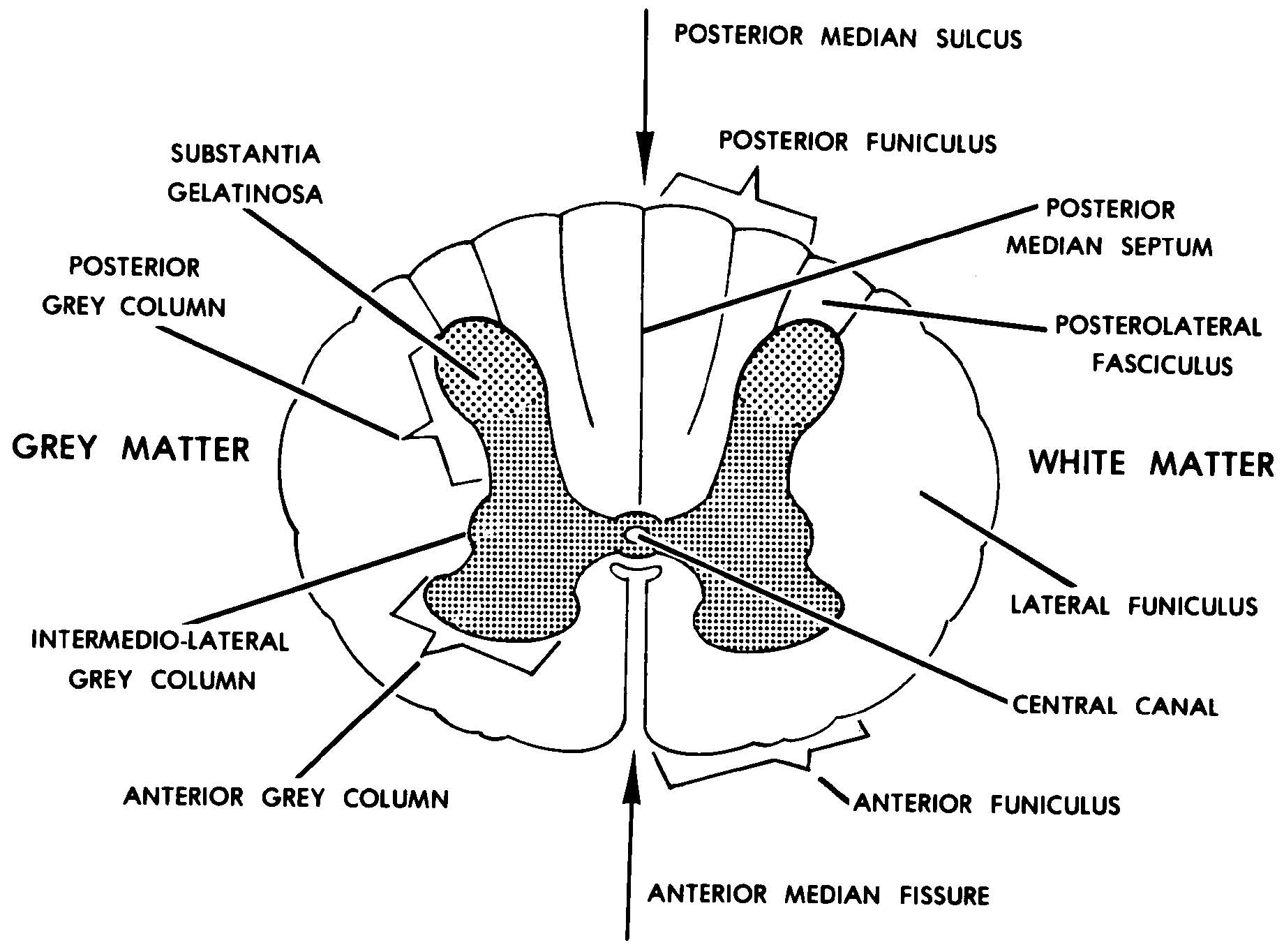
Cross Section Anatomy of the Spinal Cord. Like the brain, the spinal cord is also made up of regions of white matter and gray matter. White matter regions are comprised of axons. It appears white due to the myelin sheath on the axons. Gray matter regions are comprised of cell bodies and dendrites. Gray matter is the location of most synapses.

Operative Spinal Cord Anatomy The Neurosurgical Atlas, by Aaron CohenGadol, M.D.
Spinal Cord Cross Section Labeling by Mischief Goddess 64,431 plays 11 questions ~30 sec English 11p More 122 4.82 (you: not rated) Tries Unlimited [?] Last Played December 11, 2023 - 08:13 PM There is a printable worksheet available for download here so you can take the quiz with pen and paper. From the quiz author
Spinal Cord Anatomy Parts and Spinal Cord Functions
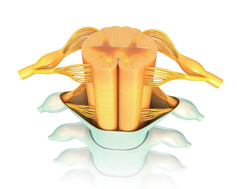
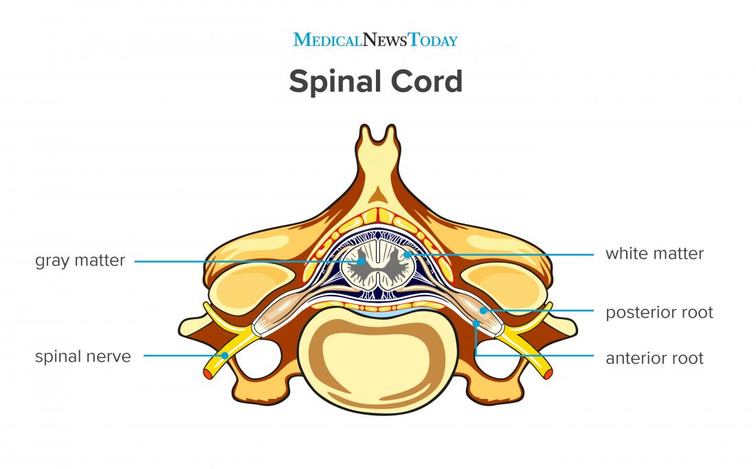
The spinal cord is a long, thin, tubular structure made up of nervous tissue that extends from the medulla oblongata in the brainstem to the lumbar region of the vertebral column (backbone) of vertebrate animals. The center of the spinal cord is hollow and contains a structure called central canal, which contains cerebrospinal fluid.
Ascending tracts of the spinal cord Anatomy Kenhub
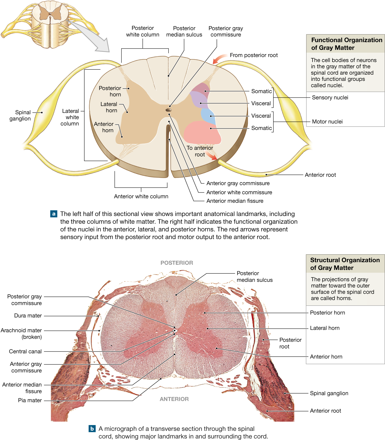
Internal anatomy of the spinal cord. The following discussion of the internal anatomy of the spinal cord will introduce some of the general principles of organization that also hold true for the brainstem. A cross-section through the spinal cord is illustrated schematically in Figure 2.6 and 3.4. The gray matter forms the interior of the spinal.

14.4 The Spinal Cord Anatomy & Physiology
Identify structures in the gross anatomy of the spinal cord on both models and cadavers or wet specimens. 3. Identify structures in the cross section of the spinal cord on classroom models. 4. Identify the nerve plexuses and specifi c nerves from each. At this point, students are responsible for the specifi c muscles innervated by each.

SpinalCordCrossSectionTractsimageVbKM.jpg 1,347×1,600 pixels Spinal cord, Spinal
Cross-sectional view of an individual spinal segment. Image by Lecturio. Cross-sectional anatomy. When viewed in cross section, the spinal cord is divided into gray matter Gray matter Region of central nervous system that appears darker in color than the other type, white matter. It is composed of neuronal cell bodies; neuropil; glial cells and capillaries but few myelinated nerve fibers.
Human Spinal Cord Crosssection Photograph by Pixologicstudio/science Photo Library
Summary The spinal cord is a long bundle of nerves and cells that extends from the lower portion of the brain to the lower back. Spinal cord functions include carrying signals between the.
PPT Chapter 13 Spinal Cord, Nerves and Reflexes PowerPoint Presentation ID735608
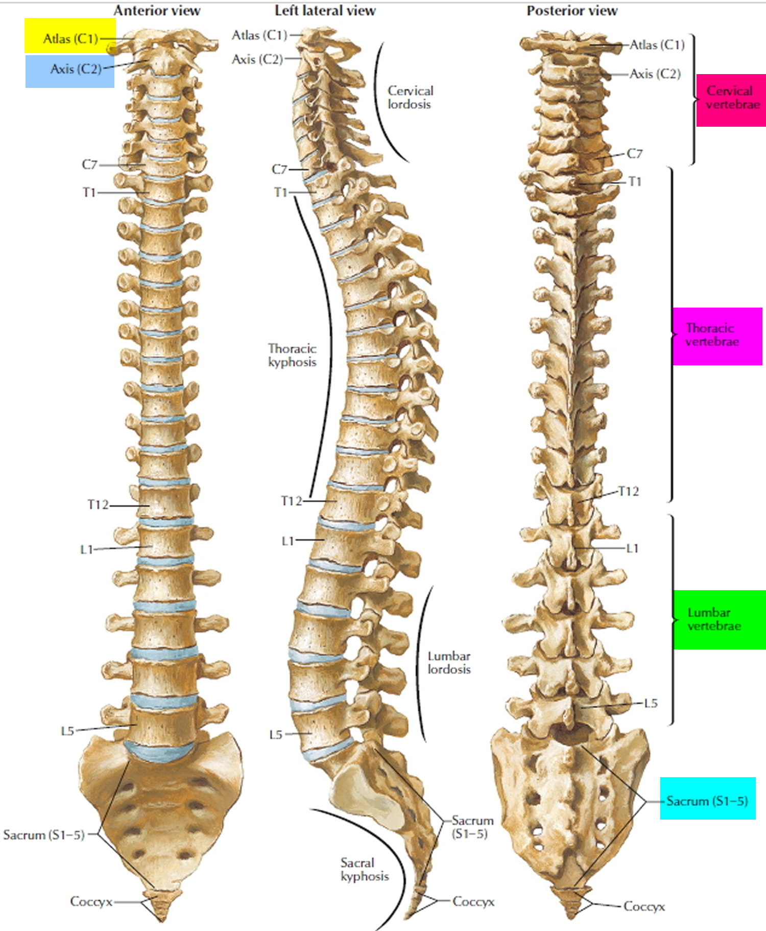
The vertebra provides several crucial functions to the body. First, it acts as a structural component of the spine, bearing body weight, anchoring muscles and the spinal cord, and forming joints with other vertebrae and ribs that allow the torso and neck to move. Second, the vertebra protects the delicate tissues of the spinal cord by.
Download Spinal Cord Gray Matter Integrates Information And Label Cross Section Of Spinal Cord
Next, the user will find anatomical sections of the spinal cord at different levels: cervical spinal cord (C2, C5), thoracic spinal cord (T10), lumbar spinal cord (L3) and sacral spinal cord (S3). Anatomy : Spinal cord, Funiculi of spinal cord, Tectospinal tract, Anterior funiculus; Ventral funiculus, Cuneate fasciculus, Gracile fasciculus.

Spinal Cord Model Bing Images Nervous system anatomy, Human anatomy and physiology, Anatomy
The spinal cord is made up of 31 segments. Each segment gives rise to a pair of spinal nerves. 1 2 In cross-section (c.s.), the segments appear to be divided into two zones. The outer zone contains many myelinated axons that run up and down the spinal cord.
Spinal Cord Anatomy Parts and Spinal Cord Functions
The spinal cord is part of the central nervous system and consists of a tightly packed column of nerve tissue that extends downwards from the brainstem through the central column of the spine. It is a relatively small bundle of tissue (weighing 35g and just about 1cm in diameter) but is crucial in facilitating our daily activities.. The spinal cord carries nerve signals from the brain to other.

Spinal Cord Diagram Cross Section Link Pico
The spinal cord is the part of the central nervous system found within the vertebral column's spinal canal. The cord extends from the corticomedullary junction at the foramen magnum of the skull down to the tip of the conus medullaris within the lumbar cistern .
Images 11. Nervous System Basic Human Anatomy
The spinal cord is a continuation of the brainstem. It extends from the foramen magnum at the base of the skull to the L1/L2 vertebra where it terminates as the conus medullaris (medullary cone).

Spinal Cord Desending Tracts at Missouri State University StudyBlue Human Anatomy Chart
Link to PayPal donation https://paypal.me/studentlamedicina?locale.x=en_UShttps://www.instagram.com/anatomy.knowledge/Spinal cord white matter organisation o.
Spinal Cord Cross Section Labeled EdrawMax EdrawMax Templates
Spinal Cord Anatomy. formed by S2, S3, S4 parasympathetic fibers and lumbar sympathetic fibers (splanchnic nerves) is residual fragment of spinal cord that extends from conus medullaris to sacrum. the dural surrounded sac that extends from the spinal cord and contains CSF, nerve roots and the cauda equina.
Spinal cord Anatomy, functions, and injuries
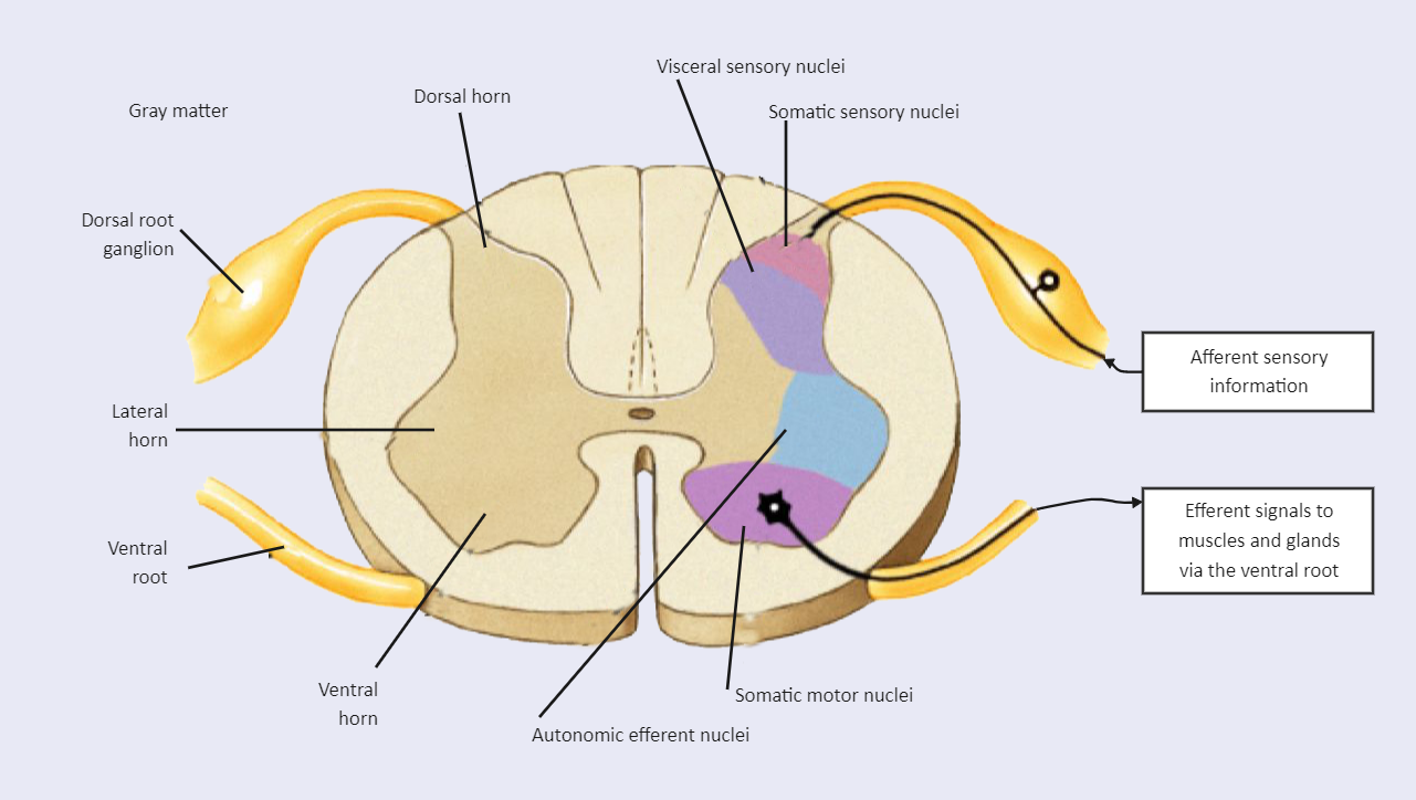
Cross section of spinal cord by Anatomy Next . Ascending tracts of spinal cord. Ascending tracts carry sensory nerve fibers from the nerve cell bodies located in the dorsal root ganglion. These pathways include the sensory information about pain, temperature, tactile sense (a sense of touch) and proprioception (perception of the position and.