
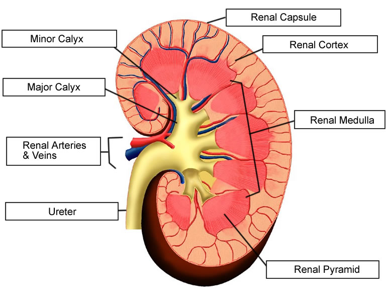
Draw the L.S of kidney and label the parts.
Kidney Anatomy and Filtration Diagram (labelled) C043/4825. Rights Managed. 27.7 MB (2.6 MB compressed) 3801 x 2550 pixels. 32.3 x 21.6 cm · 12.7 x 8.5 in (300dpi) This image is not available for purchase in your country. Please contact your Account Manager if you have any query.
5b2 Organs HumanBio
The left kidney is located at about the T12 to L3 vertebrae, whereas the right is lower due to slight displacement by the liver. Upper portions of the kidneys are somewhat protected by the eleventh and twelfth ribs (Figure 25.1.1). Each kidney weighs about 125-175 g in males and 115-155 g in females.
Human Kidney Cross Section, Scientific Background, Anatomy, Urinary System with Main Parts
Kidneys and ureters are organs of the urinary system.They take part in urine production and its transport to the urinary bladder, respectively.Fun fact is that the kidneys filter around 180 liters of blood each day, meaning that your entire blood volume passes through them around 60 times every day.. Adrenal glands (suprarenal glands) rest at the superior poles of the kidneys, but functionally.
How to Prevent and Treat Kidney Problems With Food
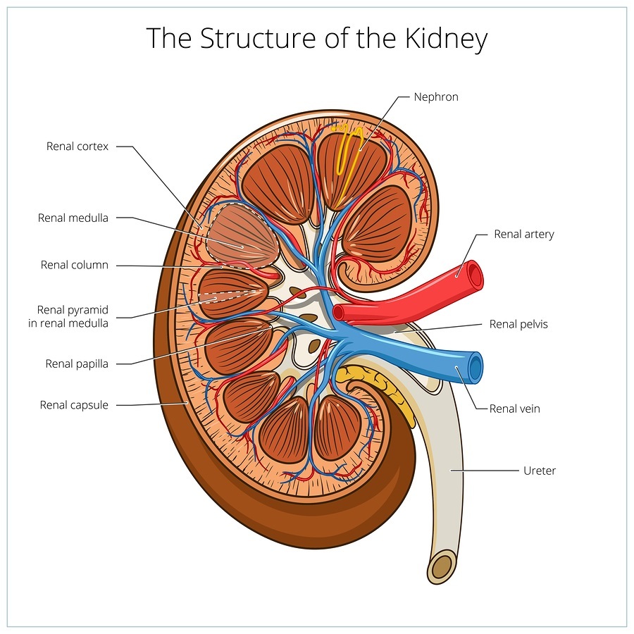
Label and Color a Diagram of the Kidney Using Listed Terms Label and Color the Kidney This worksheet has a very simplified view of a kidney showing the cortex, renal pyramids, renal artery and vein, renal pelvis, and ureter. Students can practice labeling the structures and color coding the diagram.
Labeled Kidney Anatomy Cross Section Infographic Diagram Including All Parts Stock Illustration
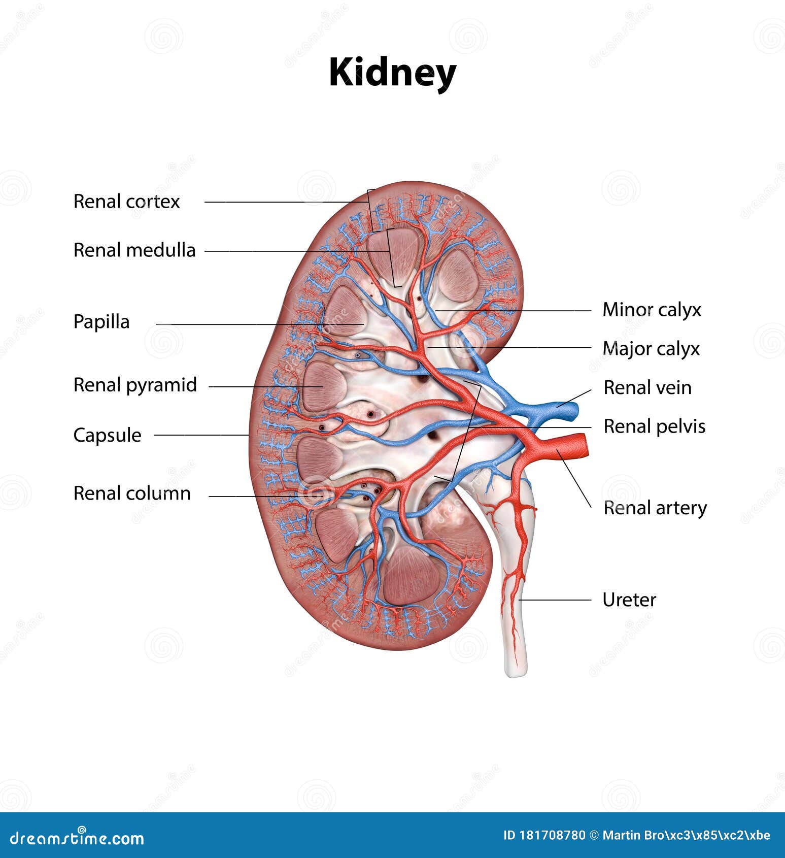
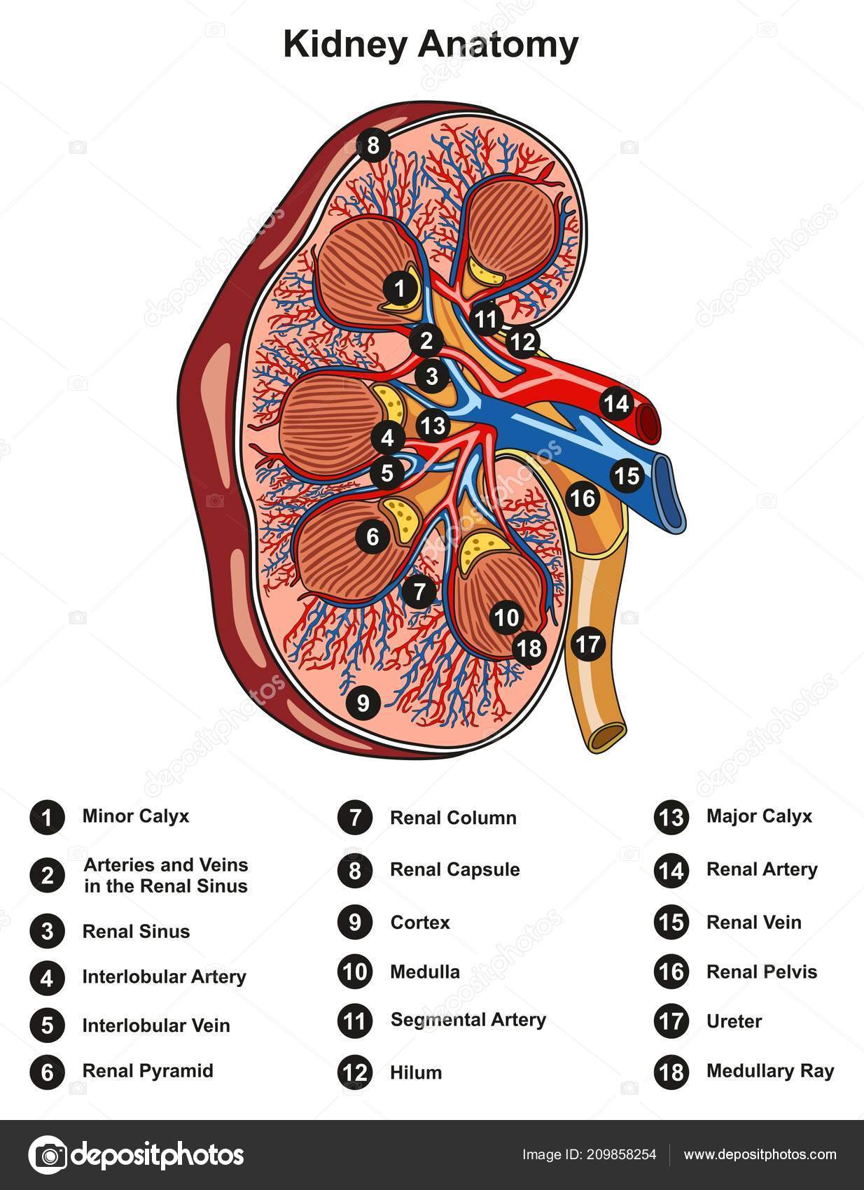
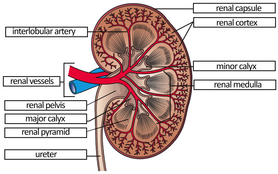
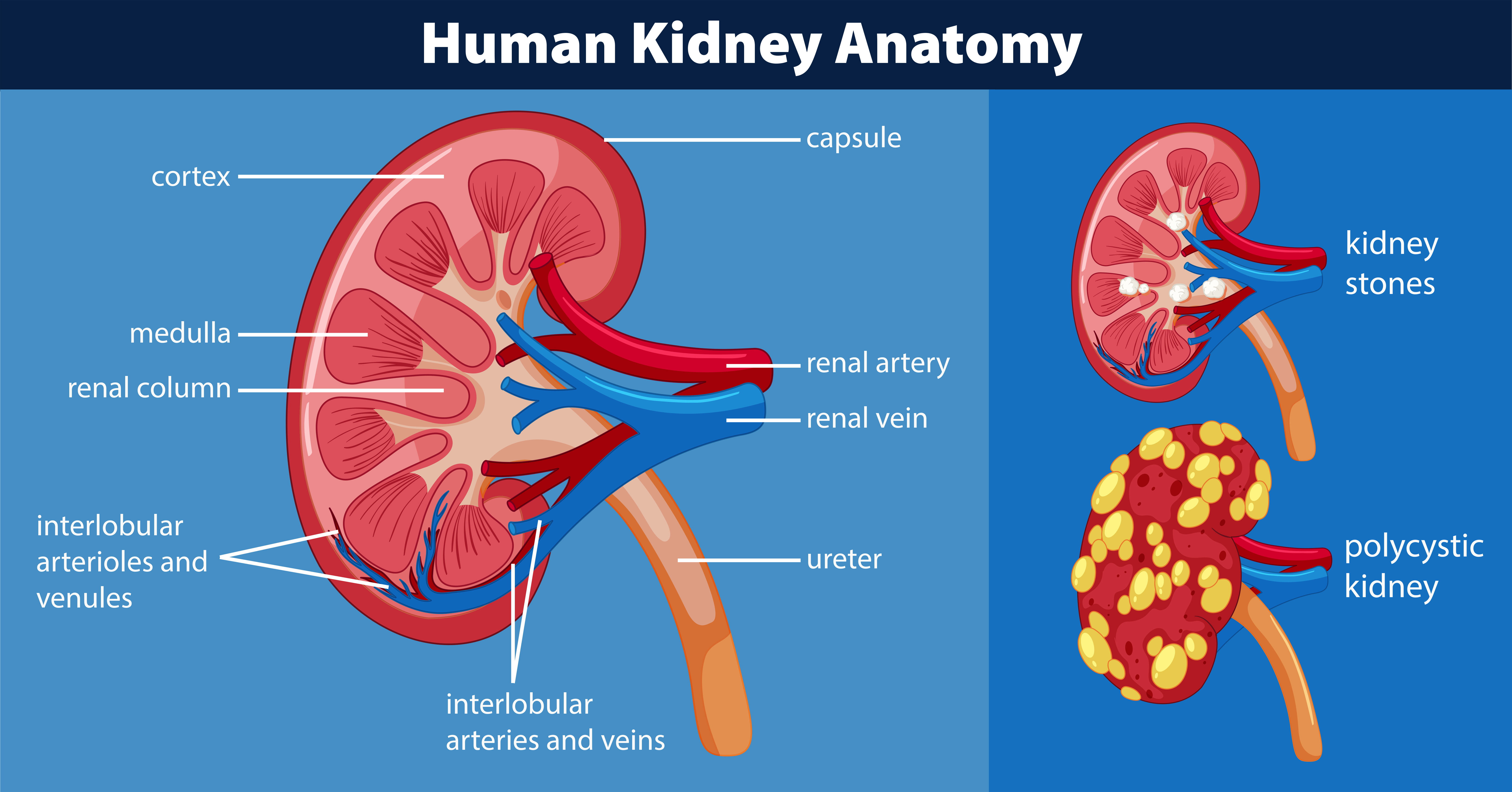
1/4 Synonyms: Cortex renalis The kidneys are paired retroperitoneal organs of the urinary system. Their function is to filter blood and produce urine. Each kidney consists of a cortex, medulla and calyces. The nephron is the main functional unit of the kidney, in charge of removing metabolic waste and excess water from the blood.
Label the Parts of the Urinary System
Quick Facts Size of an adult kidney: Length: 11-12 cm Width: 5.0-7.5 cm Weight of an adult kidney: Males: 125-170 g Females: 115-155 g Located in the abdominal cavity, kidneys are the most efficient filters. They are an important component of the human excretory system, and help the body retain essential molecules and get rid of the unwanted ones.

Normal Kidney Anatomy
That this, it is the kidney nephrons that actually perform the kidney's main functions. There are approx. a million nephrons within each kidney. To find out more about these, visit the page about kidney nephrons. Collecting Duct (Kidney): The collecting duct labelled in the diagram above is part of the kidney nephron (shown much enlarged).
Urinary System Labeling Key
1/3 Synonyms: none The kidneys are bilateral organs placed retroperitoneally in the upper left and right abdominal quadrants and are part of the urinary system. Their shape resembles a bean, where we can describe the superior and inferior poles, as well as the major convexity pointed laterally, and the minor concavity pointed medially.

What Are the Medullary Pyramids? (with pictures)
The nephron is the small function unit of the kidney. It consists of a renal corpuscle and a renal tubule. The renal corpuscle has a saclike structure - the glomerular capsule - which contains a tuft of blood capillaries, called the glomerulus. The blood capillaries in the glomerulus filter fluid, which is the first step in urine formation.
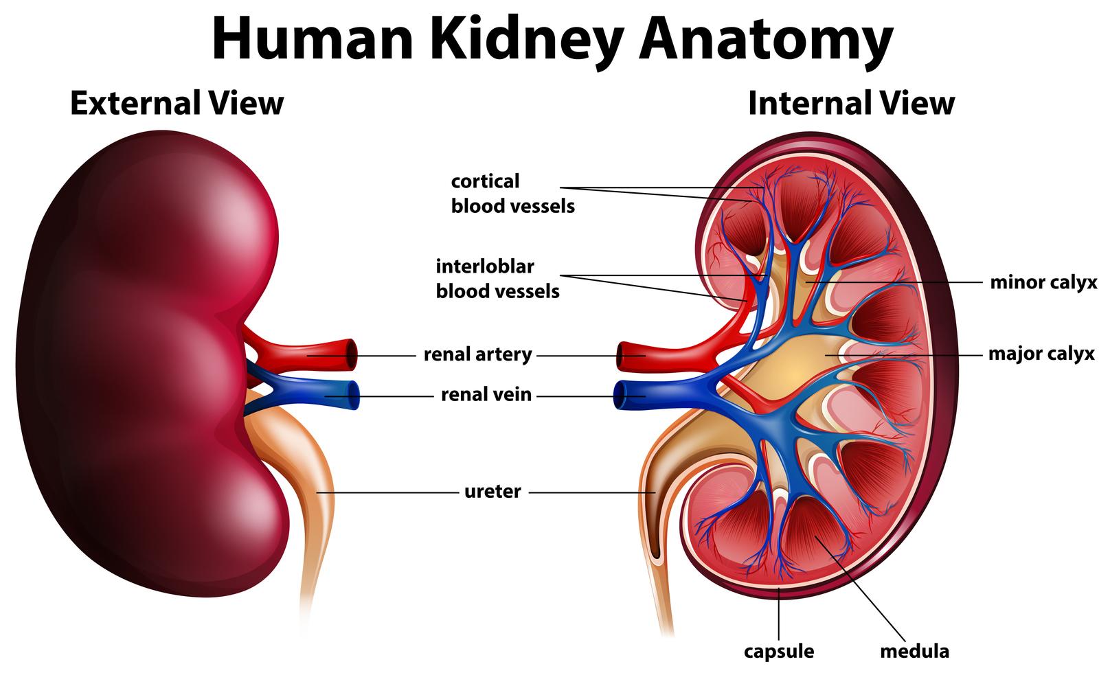
Diagram showing human kidney anatomy 295196 Vector Art at Vecteezy
Updated on April 06, 2022 Medically reviewed by Isabel Casimiro, MD, PhD Table of Contents Anatomy Function Associated Conditions Tests Treatment The kidneys are the body's filtration system. These fist-sized, bean-shaped organs manage the body's fluid and electrolyte balance, filter blood, remove waste, and regulate hormones.
Human kidney anatomy diagram 434204 Vector Art at Vecteezy
In (a), the large cell body can be seen at the top right corner, with branches extending from the cell body. The smallest finger-like extensions are the pedicels. Pedicels on one podocyte always interdigitate with the pedicels of another podocyte. (b) This capillary has three podocytes wrapped around it.

Human kidney medical diagram with a cross section Vector Image
What are kidneys? The kidneys are two bean-shaped organs in the renal system. They help the body pass waste as urine. They also help filter blood before sending it back to the heart. The.
What Are the Parts of the Human Kidney? Healthfully
The kidneys are two bean-shaped organs that filter your blood. Your kidneys are part of your urinary system. Your kidneys filter about 200 quarts of fluid every day — enough to fill a large bathtub. During this process, your kidneys remove waste, which leaves your body as urine (pee). Most people pee about two quarts daily.

Please send me a diagram of L.S Of kidney and label the main parts of it.. Brainly.in
Below is a well-labelled and easy diagram of the nephron for your better understanding. Broadly, a nephron can be divided into two parts - renal corpuscle and renal tubule. The renal corpuscle is the filtering component and renal tubules carry the filtered liquid away. A human nephron is a long fine tubule that is about 30-55 mm long.

Diagram Of Human Kidney Anatomy Stock Illustration Download Image Now iStock
Each kidney weighs about 125-175 g in males and 115-155 g in females. They are about 11-14 cm in length, 6 cm wide, and 4 cm thick, and are directly covered by a fibrous capsule composed of dense, irregular connective tissue that helps to hold their shape and protect them.
Human kidney anatomy diagram 446409 Vector Art at Vecteezy
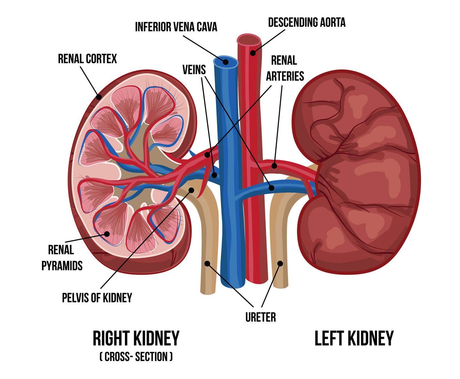
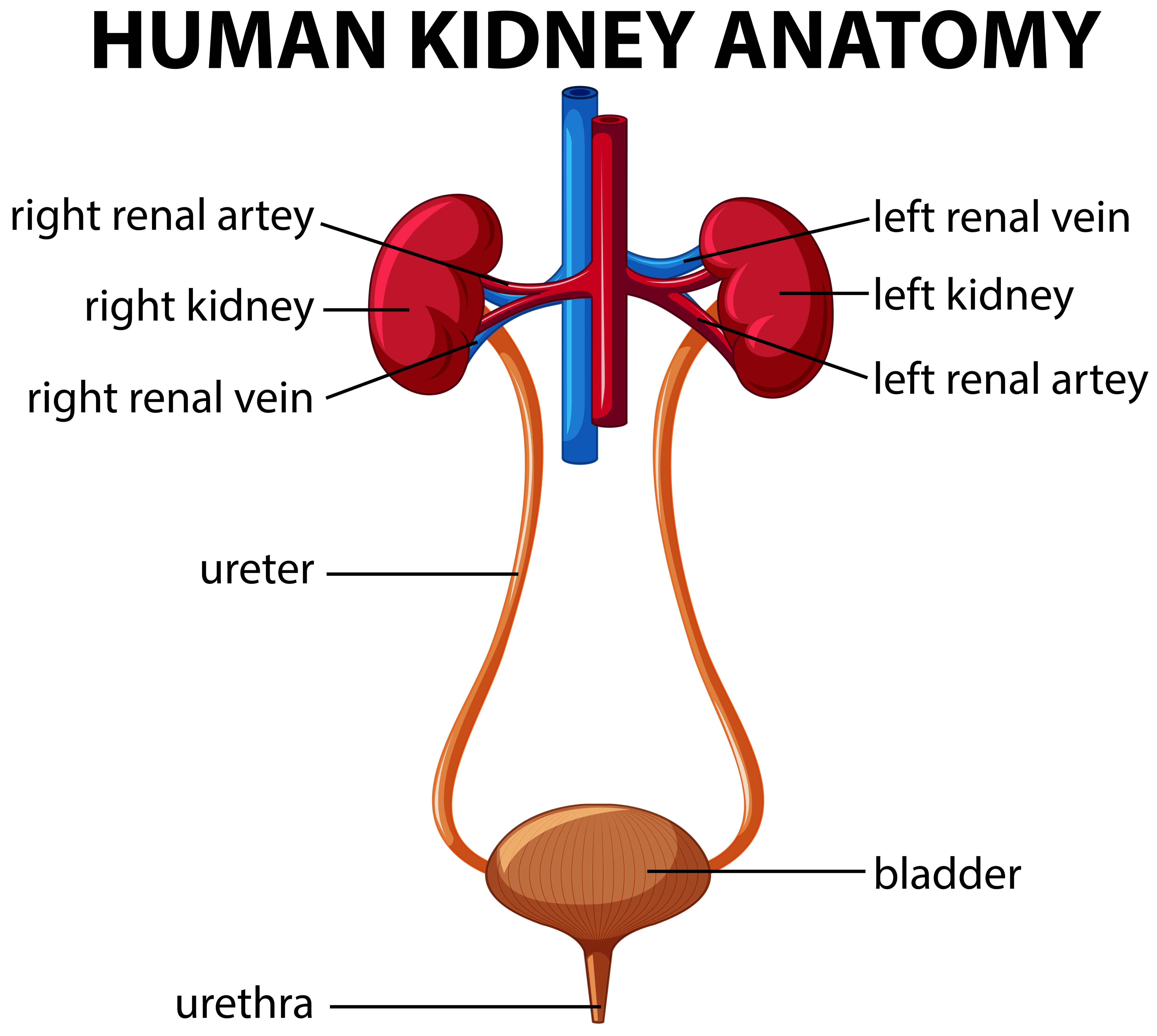
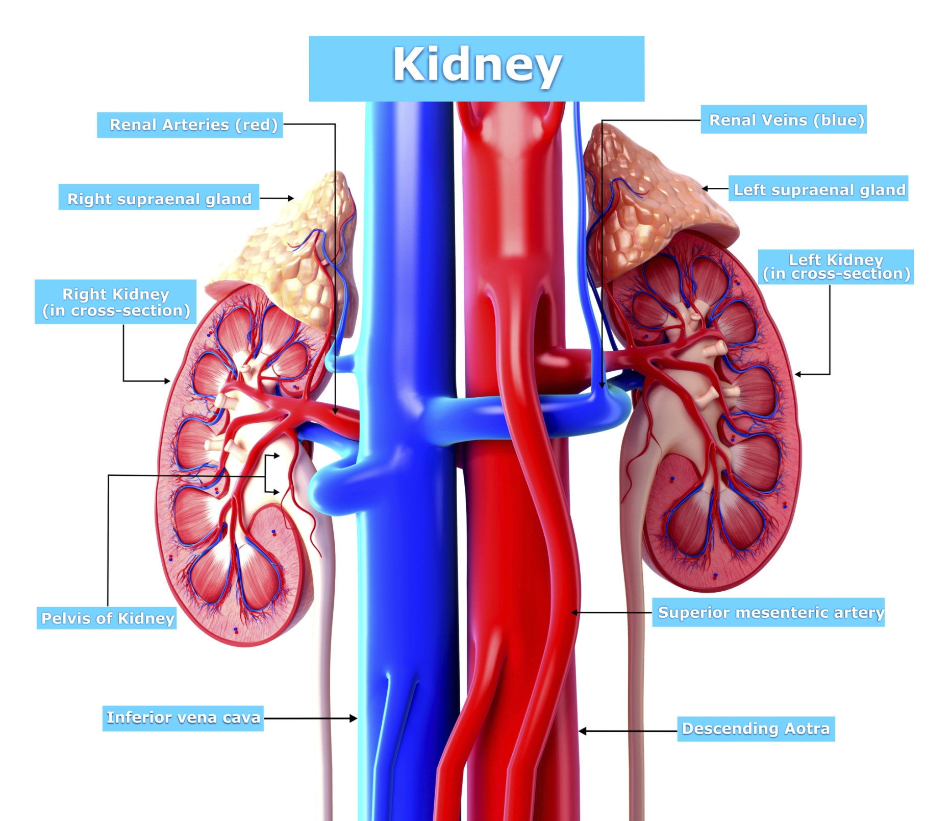
The first slide is an overview of the urinary system that shows the kidneys, ureters, urinary bladder, and urethra. Students drag labels to the structures on the slide. Also, the diagram shows the relationship between the aorta, vena cava, and the renal vessels.